| Home Page
Case Presentation
Various Instruments.
Contact Page
Custom Page
Custom2 Page
Custom3 Page
Custom4 Page
Favorite Links Page
Guest Book Page
Cases
Tonsillectomy
SMR
INA and Caldwell Luc
Mastoid operations
Radiology
Shopping Page Page
Slide Show Page
Whats New Page
|
|
Sinus Disease
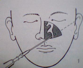
If despite antibiotic therapy the patient continues to have sinus disease,then a proof puncture is made to either get a pus for culture and sensitivity of to proove that indeed there is sinus disease since radiological images can be inconclusive. Here after anesthtising the nose with cottonoids soaked in 4% xylocaine and inserted into the infrerior meatus, the Tilley Lichtwicz trochar with the canula inserted is introduced into the nose into the inferioir meatus and with a boring action the lateral nasal wall is puntured and the antrum gained access.Then the trochar is removed and a 10cc syringe attached to the canula and the returning pus is extracted and transferred into a sterile bottle to be sent for culture and sensitivity.
The antrum is washed with sterile saline by attaching the canula to a Higginsons syringe and by compressing the bulb of the syringe, saline is fluched into the sinus and the returning fluid is made to collect into a basin by asking the patient to bend over one .
|
|
|
Antral Puncture
Care must be taken to direct the trochar to the ipsilateral tragus...otherwise if directed superiorily, the orbit can be puntured or if directed laterally , the lateral antral wall can be punctured through and through and the cheek can bulge when saline is injected.
|
|
|
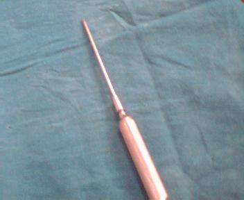
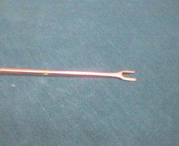
Tilley Lichtwicz trochar canula
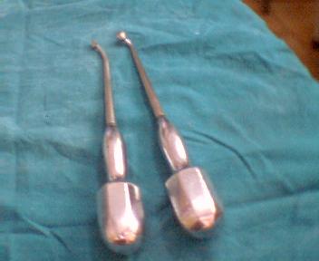
If inspite if antral puncture , the maxillary antrum continues to be diseased , then a permanent draining window is made in the inferior meatus, 1/2 and inch behind the anterior end of the inferior meatus, where the inferior turbinate curves backwards, a point called the genu,where,the pertition wall between the inferior meatus and the antrum is thinnest.This window is made using the tilley's antral harpoon, held like a dagger and with force the wall broken open.The ragged edges of this opening is made smooth using the tilley's antral burr.This procedure is called Intra Nasal Antrostomy or INA.
As a result, the antrum was expected to drain by gravity...today we know that the antrum only drains by mucociliary clearance , by the mechanism of action of the cilea towards the natural ostium, located high up in the middle meatus.There the opening made by INA was nonfuntional.
Now, a functional opening is made in the middle meatus using an endoscope an by inserting the Stambergers rhinoforce reverse cutting forceps into the accessory ostium located below and behind the bulla and cutting in a forward direction to meet and connect with the natural ostium and create a wide opening.These two ostia are connected to prevent the circus phenomenon..wherein , mucous will come out of the natural and go back into the antrum through the accesssory ostium.This opening made is called Middle Meatal Antrotomy or Supra Turbinal Fenestration or, since now it is a functiona opening, called, Functional Endoscopic Sinus Surgery, a term coined by David Kennedy of USA.
|
|
|
Tilley's antral harpoon and tilley's rosettes antral burr
This is my good friend Hal. I took this picture on his birthday. I think he likes to be in pictures. |
|
|
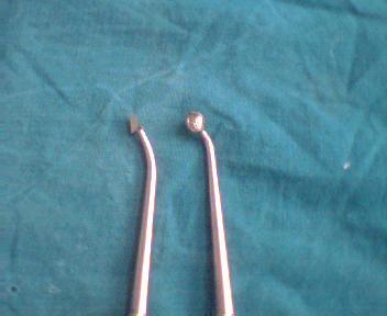
Close up of tilley's antral harpoon and antral burr
This is my good friend Hal. I took this picture on his birthday. I think he likes to be in pictures.
|
|
|
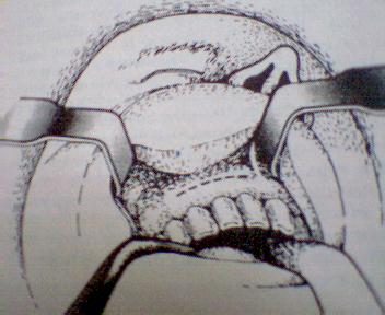
Intra Nasal Antrostomy...the free communicating opening between the inferior meatus and the maxillary antrum
The antrum was presumed to drain by gravity into the nose..but today we know that the drainage is to the natural ostium located high up in the middle meatus, seen above the inferior turbinate in the picture. |
|
|
The Caldwell Luc Operation
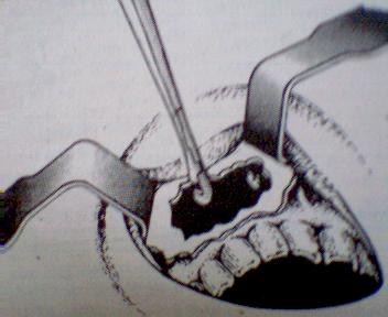
If despite an INA the antrum is grossly diseased, then the antral contents cowuld have to be cleared by a procedure called radical antrotomy or the Callwell Luc Operation in which the patient is placed semi-sitting with the head flexed onto the trunk , the area of the upper lip prepared and, the patient draped.
A curvilinear incision is made just below the gingivo labial Sulcus starting from the canine tooth using a 10 no knife and extending laterally across the canine fossa. The periosteum is elevated using the periosteal elevator and and at the depth of the canine fossa , using a gouge the partition wall is broken and the antrum gained access. The diseased antral contents are curetted out and intranasal antrostomy performed. The antrum is packed with medicated ribbon gauze, whose end is brought out through the INA opening and out through the nose. Suturing the incision site closes the opening made. The packing is removed after 48 hours.
|
|
|
Making the opening into the antrum using a gouge
|
|
|