Radiology Page
     
Home Page

Case Presentation

Various Instruments.

Contact Page

Custom Page

Custom2 Page

Custom3 Page

Custom4 Page

Favorite Links Page

Guest Book Page

Cases

Tonsillectomy

SMR

INA and Caldwell Luc

Mastoid operations

Radiology

Shopping Page Page

Slide Show Page

Whats New Page


Plain x-ray waters view shows an air fluid level in the right maxillary sinus .Maybe caused by acute sinusitis, sinus barotrauma, or trauma.Tilting the head of the patient and taking a repeat radiograph may confirm the presence of fluid.


Foriegn body in the upper oesophagus..from its shape presumably a coin.To distuinguish, take a lateral xray...will show it is lying in the upper oesophagus and not in the trachea.

this has to be removed using a hypopharyngoscope under GA.


Retropharyngeal abscess

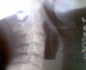
A retropharyngeal abscess is inferred when the prevertebral soft tissue shadow is more than 2/3 rd the thickness of the body of the corresponding vertebrae.


 RRPA develops secondary to lymphatic drainage or contiguous spread of upper respiratory or oral infections. Pharyngeal trauma from endotracheal intubation, endoscopy, foreign body ingestion, and removal may cause a subsequent RPA. Patients who are immunocompromised or chronically ill, such as persons with diabetes, cancer, alcoholism, or AIDS, are at increased risk for RPA.

The most common organisms causing retropharyngeal abscesses include aerobes and anaerobes; gram-negative organisms also may be observed. Often, mixed flora are cultured.

Retropharyngeal abscess generally affects children under age 5. Tissues at the back of the throat in young children allow a pus-filled space to form immediately behind the back of the throat. This area can become secondarily infected during or immediately following a bacterial sore throat.

The affected child, who may still have symptoms of the original sore throat, develops a high fever with an extremely severe sore throat. The pain causes difficulty swallowing and the expanding abscess may interfere with breathing. Complications can be life-threatening.

Retropharyngeal abscess requires immediate attention to prevent severe complications. Surgical drainage of the abscess and high-dose intravenous antibiotics are used to treat the infection. The airway needs to be protected from becoming completely blocked by the swelling.

Complications:

  • Airway obstruction
  • Mediastinitis
  • Pleural involvement
  • Atlantooccipital dislocation
  • Epidural abscess
  • Sepsis
  • Adult respiratory distress syndrome (ARDS)
  • Erosion of the second and third cervical vertebrae
  • Cranial nerve deficits (cranial nerves IX-XII are contained in the cervical fascia)
  • Septic thrombosis of jugular vein or hemorrhage secondary to erosion into carotid artery.

Irregular filling defect in the lower third of the oesophagus..suggestive of carcinoma oesophagus

Oesophageal carcinoma

  • 90% are squamous cell carcinomas

  • Occur in the upper or middle third of the oesophagus

  • 8% are adenocarcinomas
  • Occur in the lower third of the oesophagus
  • Overall 5 year survival is very poor and is at best 20%
  • Less than 50% patients are suitable for potentially curative treatment
  • Of those undergoing 'curative' treatment less than 40% survive one year

Risk factors

  • Squamous cell carcinoma
    • Alcohol / tobacco
    • Diet high in nitrosamines
    • Aflatoxins
    • Trace element deficiency - molybdenum
    • Vitamin deficiencies - vitamins A & C
    • Achalasia
    • Coeliac Disease
    • Genetic - Tylosis
    • High incidence in Transkei, Areas of Northern China and the Caspian littoral region
  • Adenocarcinoma
    • 15% associated with Barrett's Oesophagus

Clinical features

  • Progressive dysphagia
  • Respiratory symptoms due to overspill or occasionally a trachea-oesophageal fistula
  • Weight loss

Assessment

Diagnosis confirmed by:

  • Endoscopy plus biopsy / cytology
  • Barium swallow

oesophageal carcinoma

Resectability and fitness for surgery assessed by:

  • Chest x-ray
  • Lung function tests(FEV1 > 1L)
  • Liver ultrasound
  • Endoscopic ultrasound
  • Bronchoscopy
  • Laparoscopy
  • Thoracic CT

Management

  • Adenocarcinomas are not radiosensitive and surgery is mainstay of treatment
  • Squamous cell carcinomas can be treated with either surgery or radiotherapy

Radiotherapy

  • Pearson in Edinburgh (1977)
  • 19% 5 years survival with radiotherapy
  • Improved survival compared to surgery
  • Similar results not seen in other centres

Surgery

  • Only 40% tumours are resectable
  • Operative mortality now less than 10%
  • Treatment should be in centres who perform operation regularly
  • No place for the occasional operator
  • Preoperative chemotherapy may be beneficial

Operative approaches

Need 10 cm proximal clearance to avoid submucosal spread.

  • Total gastrectomy via thoracoabdominal approach (Adenocarcinoma)
  • Subtotal two-stage oesophagectomy (Ivor-Lewis)
  • Subtotal three-stage oesophagectomy (McKeown)
  • Transhiatal oesophagectomy

Palliative treatment

Aim to relieve obstruction and dysphagia with minimal morbidity

  • Oesophageal intubation
    • Open surgical intubation (Celestin or Mousseau-Barbin tubes) now obsolete
    • Endoscopic or radiological placement now most commonly practiced
    • Atkinson tube is the most commonly placed endoscopically
    • Requires dilatation with risk of oesophageal perforation
    • Recent increased use of self-expanding stents that require no pre-dilatation
    • Complications of stents and tubes:
      • Oesophageal perforation
      • Tube displacement or migration
      • Tube blockage due to ingrowth or overgrowth

Oesophageal tubes

  • Laser therapy
    • Produces good palliation in over 60% of cases
    • May need to be repeated every 4 to 6 weeks
    • Associated with oesophageal perforation in about 5% cases
  • External beam radiotherapy
  • Brachytherapy
  • Diathermy
  • Alcohol injection

This is one of my favorite images
Place anything like pictures, sketches, logos or products on this page!

This is one of my favorite images
I took it when he wasn't looking. I took it when he wasn't looking. I took it when he wasn't looking. I took it when he wasn't looking. I took it when he wasn't looking.

This is one of my favorite images
Place anything like pictures, sketches, logos or products on this page!